
Interview with Dr Ajaya Jha
When IDG learnt that India has the unique distinction of having Asia's FIRST (& world's THIRD) intra operative MRI known as Brain Suite installed, we decided to visit Max Super Speciality hospital at Saket, New Delhi and meet up with Dr Ajaya Jha - Director, Neurosurgery.
IDG - Dr Jha, what prompted MAX Super specialty hospital to go in for Brain Suite?
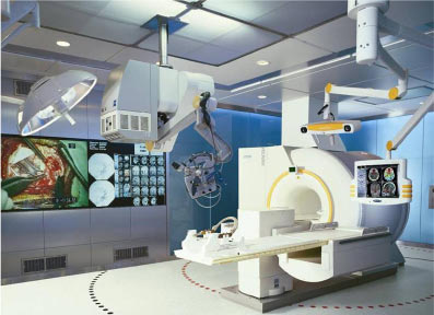
Dr Ajaya Jha - It was essentially the technology of intra operative MRI that promises to cut brain tumour patients' risks and prolongs their lives.
This comprehensive system allows brain-tumor patients to be rotated into a magnetic resonance imaging machine so doctors can see real time images while operating and therefore get an accurate look at how much residual tumor remains before ending surgery.
IDG - that means tumour tissue being left out and second time surgeries will be avoided.
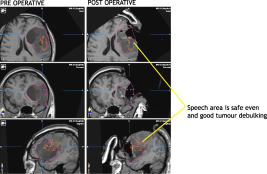
Dr Ajaya Jha - Not only that, Brain Suite even improves the accuracy manifold & hence maximizes the chances of removing brain tumors as completely as possible as well as safe guarding the critical areas like the speech area etc.
IDG - How many cases have benefited from Brain Suite till date
Dr Ajaya Jha - the first surgery was performed on Mr. PN, a British national on May 13, 2006. The patient was discharged on May 17, 2006 after a successful surgery. The team has performed "Twenty Eight Successful Surgeries" since then using the Brain Suite.
IDG - Dr Jha, can we take a picture for the benefit of IDGians?
IDG - Dr Jha, please narrate a case for our members to understand the technology better.
Dr Ajaya Jha -
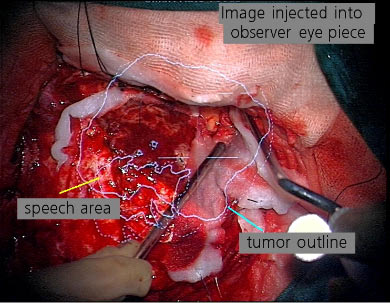
A 44 yr old gentleman underwent a biopsy abroad for a tumour which was very close to his speech area. He came to us because his tumour had increased in size and he wanted his speech to be intact after surgery. We did an MRI before surgery, to map out his speech area and using the navigation computer were able to see the tumour in relation to this. At surgery it is not possible to make out these areas as the tumor tissue looks very similar to normal brain. In our operation theatre, with the latest image guidance and microscope the speech area and tumour area were on the surgeon's eye piece and hence when the tumour was being removed we could stay well away from the speech area and got a good resection keeping his speech intact.
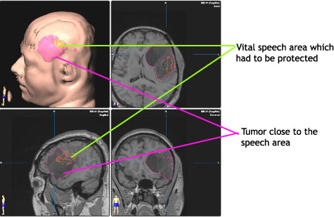
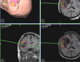
Image as seen in Microscope: The speech area is not different from tumour and because of planning with the help of navigation, the surgeon removes only tumour.
Read More: Case of Diabetes insipidus due to a suprasellar tumour